CMF Implants Made Of PEEK Polymer
PEEK-OPTIMA™ polymers solutions for strong, permanent implants following trauma
Why PEEK is an Excellent Solution for CMF Implants
Permanent, low profile PEEK solutions for craniomaxillofacial surgery reconstructions
fast fact
PEEK is cost-effective
Clinical studies show that choosing PEEK means you are 3X less likely to need to pay for a revision2 and the implants are less likely to fail3
fast fact
A more natural, lightweight feel
PEEK-OPTIMA polymers have a modulus similar to natural bone, and is lighter weight and more comfortable than metals4-8
fast fact
lmproved aesthetic & wound healing
Compared to metal, PEEK CMF implants are less likely to result in problems with healing, and are less visible under the skin1,2

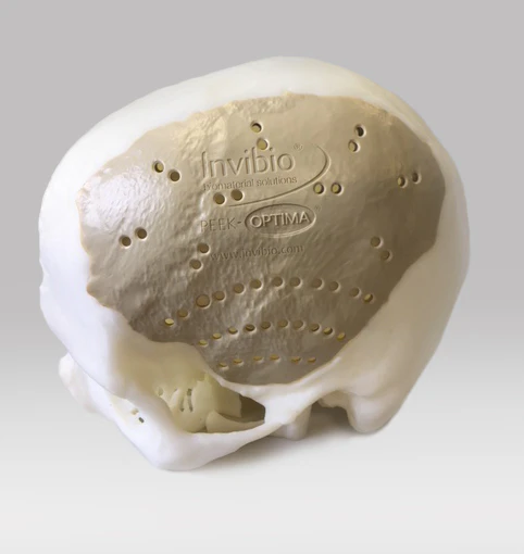
Patient-specific PEEK polymer CMF implants
PEEK-OPTIMATM high performance polymer is a strong, permanent, functional alternative to traditional structural materials like metal that can be used to manufacture custom cranial implants. PEEK-OPTIMA’s biomechanical properties are similar to bone, result in better clinical outcomes and have improved cosmetic satisfaction compared to titanium1-9.
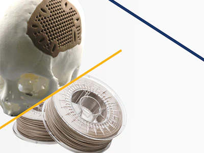
Unlocking the possibilities for 3D printing PEEK structures
We are at the fore of innovation, working with industry pioneers to meet unrealised needs
press release
A new implantable PEEK polymer form optimized for 3D printing
Invibio launches PEEK-OPTIMA™ AM filament, making available the trusted implantable-grade polymer in a form specifically developed for Fused Deposition Modeling (FDM) and Fused Filament Fabrication (FFF) additive manufacturing processes.
video
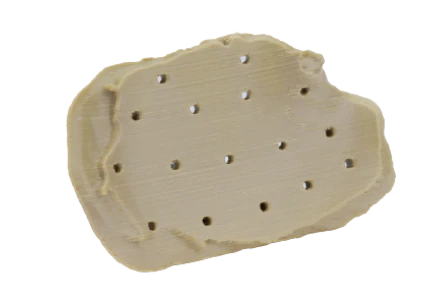
Bond 3D technology enables additive manufacture of functional parts with isotropic strength using existing PEEK-OPTIMATM Natural producing parts that retain excellent mechanical characteristics.
ARTICLE
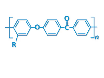
The molecular makeup of PAEK, PEKK and PEEK polymers
Chief Scientist John Grasmeder explains the unique physical properties of PAEK at the molecular level.